Autoimmune disorders are diseases in which inherent protection systems mount an attack against self-antigens. In a normally functioning immune system foreign invaders are attacked, destroyed, and eliminated. To do this, the immune system must recognize self from non-self and limit the response to foreign antigens. In diseases such as RA, the immune system fails to make this distinction and launches an attack against the synovium, the interior lining of the joint.
Purpose
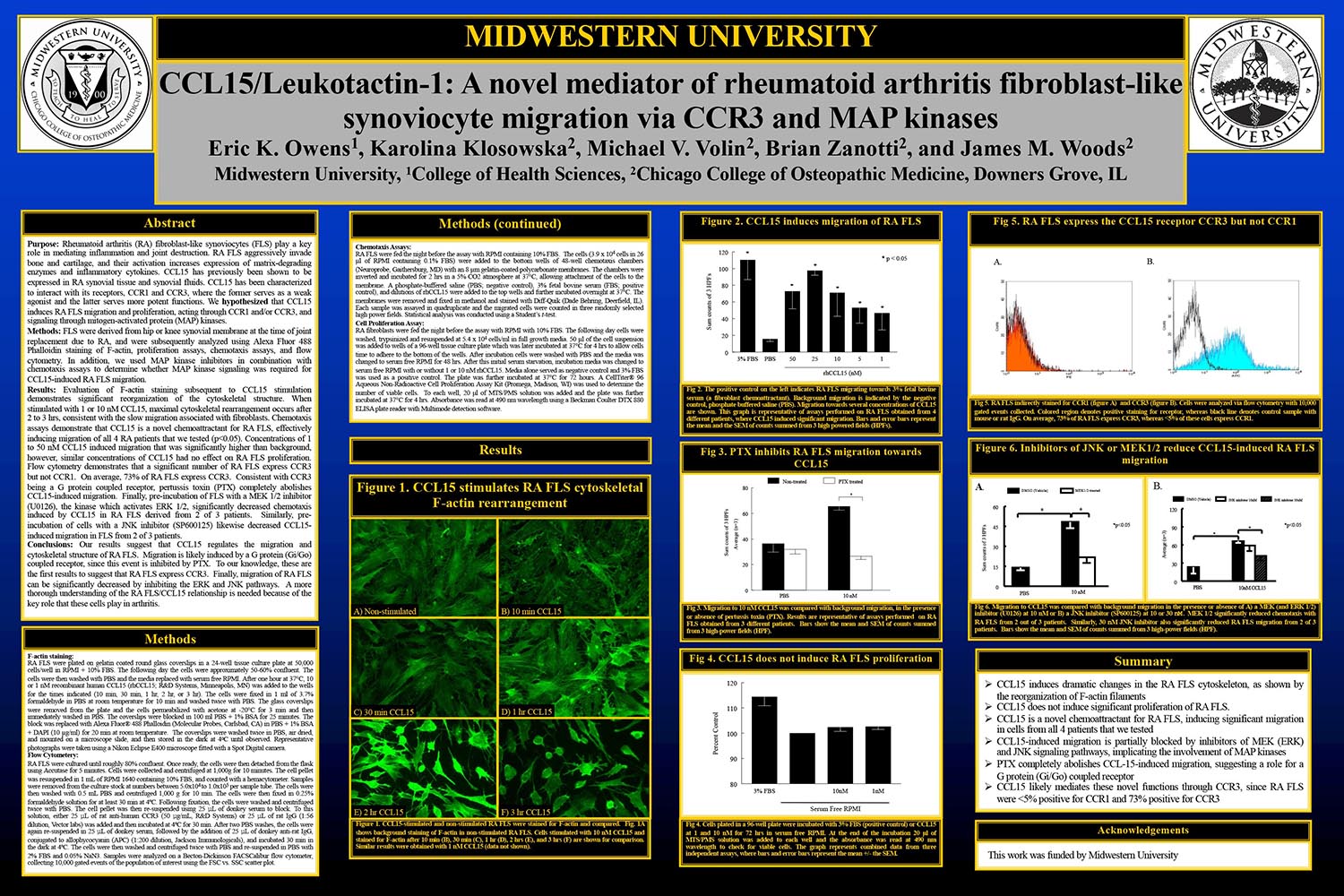
Rheumatoid arthritis (RA) fibroblast-like synoviocytes (FLS) play a key role in mediating inflammation and joint destruction. RA FLS aggressively invade bone and cartilage, and their activation increases expression of matrix-degrading enzymes and inflammatory cytokines. CCL15 has previously been shown to be expressed in RA synovial tissue and synovial fluids. CCL15 has been characterized to interact with its receptors, CCR1 and CCR3, where the former serves as a weak agonist and the latter serves more potent functions. We hypothesized that CCL15 induces RA FLS migration and proliferation, acting through CCR1 and/or CCR3, and signaling through mitogen-activated protein (MAP) kinases.
Methods
FLS were derived from hip or knee synovial membrane at the time of joint replacement due to RA, and were subsequently analyzed using Alexa Fluor 488 Phalloidin staining of F-actin, proliferation assays, chemotaxis assays, and flow cytometry. In addition, we used MAP kinase inhibitors in combination with chemotaxis assays to determine whether MAP kinase signaling was required for CCL15-induced RA FLS migration.
Results
Evaluation of F-actin staining subsequent to CCL15 stimulation demonstrates significant reorganization of the cytoskeletal structure. When stimulated with 1 or 10 nM CCL15, maximal cytoskeletal rearrangement occurs after2 to 3 hrs, consistent with the slow migration associated with fibroblasts. Chemotaxis assays demonstrate that CCL15 is a novel chemoattractant for RA FLS, effectively inducing migration of all 4 RA patients that we tested (p<0.05). Concentrations of 1 to 50 nM CCL15 induced migration that was significantly higher than background, however, similar concentrations of CCL15 had no effect on RA FLS proliferation. Flow cytometry demonstrates that a significant number of RA FLS express CCR3 but not CCR1. On average, 73% of RA FLS express CCR3. Consistent with CCR3 being a G protein coupled receptor, pertussis toxin (PTX) completely abolishes CCL15-induced migration. Finally, pre-incubation of FLS with a MEK 1/2 inhibitor (U0126), the kinase which activates ERK 1/2, significantly decreased chemotaxis induced by CCL15 in RA FLS derived from 2 of 3 patients. Similarly, pre-incubation of cells with a JNK inhibitor (SP600125) likewise decreased CCL15-induced migration in FLS from 2 of 3 patients.
Conclusions
Our results suggest that CCL15 regulates the migration and cytoskeletal structure of RA FLS. Migration is likely induced by a G protein (Gi/Go) coupled receptor, since this event is inhibited by PTX. To our knowledge, these are the first results to suggest that RA FLS express CCR3. Finally, migration of RA FLS can be significantly decreased by inhibiting the ERK and JNK pathways. A more thorough understanding of the RA FLS/CCL15 relationship is needed because of the key role that these cells play in arthritis.
Download the rest of the whitepaper here.
- Water, IV Hydration And The Implications For Tight Muscles - July 15, 2019
- The Collagen Craze: How Collagen Peptides Differ From The Collagen Causing Your Muscle Pain - June 28, 2019
- Anatomy Trains, Collagen and the Delos Perspective - September 20, 2018


